- Intralase Lasik – Custom Bladeless Lasik
- Avedro Crosslinking – Lasik-Xtra
- Refractive Lens Exchange
Approved by NASA Astronauts and NAVY pilots
3 reasons to choose us for your LASIK surgery:
- We only use disposable instruments during your surgery.
- Your annual follow-up consultation is included in the price of your procedure. If you miss an appointment one year we still honour it the following year, while with other providers in this case you lose the warranty.
- We include a Pentacam / Optos exam before your surgery ($400 value) completely free to ensure the best results.


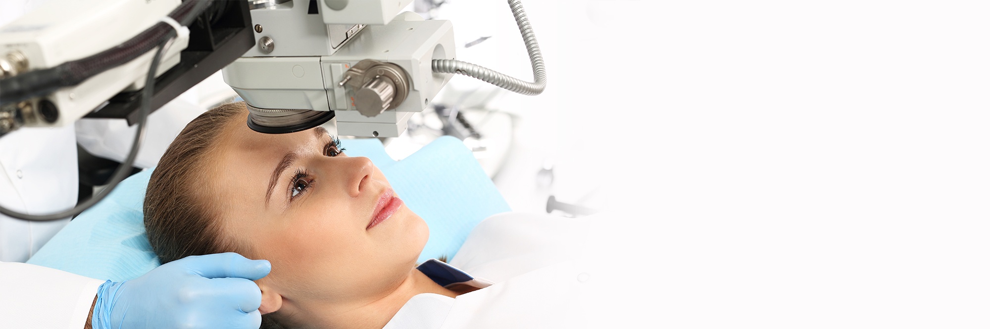
During LASIK surgery, a flap is created in the outer layers of the cornea. The flap is hinged and lifted so the excimer laser can treat only the underlying layer of the cornea. This circular flap remains attached to the cornea by a small hinge of tissue. The hinge enables the flap to be lifted away from the central cornea. A laser can then be used to reshape the exposed mid-layer of the cornea.
The IntraLase® laser is used to create a flap using multiple short pulses. These pulses are so close together they create an almost complete separation of the flap from the rest of the cornea, but they do not actually lift the flap. When the flap pattern is judged to be complete and satisfactory, a delicate separation of the flap is performed with a few gentle manipulations using a surgical instrument.
While creating the flap, the eye is held firmly with a suction ring, which exerts some pressure and causes vision to black out momentarily.
The surgeon then positions the patient’s eye under the excimer laser which is programmed to remove microscopic layers of tissue from the internal part of the cornea under the flap. The cool laser beam vapourizes tissue away, one microscopic layer at a time, without burning or cutting. This tissue does not completely replace itself after it is removed. Since the excimer laser light is created at a specified wavelength that does not pass through the cornea, no other part of the eye is affected.
After the tissue has been removed, the surgeon places the flap back in its original position where it heals into place without the necessity of stitches. The cornea has amazing natural bonding qualities. Within a few minutes, the flap adheres to the underlying tissue. The edges of the flap heal over in 12 to 48 hours, with the entire flap gaining adhesive strength as it continues to heal in the following weeks and months.
For each eye, the laser application time is usually less than one minute and the whole procedure takes around 15 minutes.
Watch our video testimonial
iLASIK Fact Sheet
For more information, email lasik@patodiaeye.com
Corneal Collagen Cross-Linking (KXL) is a surgical procedure used to strengthen a weakened cornea. The most common cause of a weakened cornea is called Keratoconus.
LASIK-Xtra™ is an addition to the normal LASIK procedure which is designed to minimize the weakening effects of LASIK on the cornea by strengthening it with cross-linking treatment. Corneas normally become thinner after LASIK because the excimer laser used removes tissue and especially after Standard or Custom LASIK is performed with a blade that creates a much thicker flap than a blade-free procedure.
This effect is more marked when higher degrees of spectacle power are treated. In a small percentage of cases, this thinning causes the cornea to bulge forwards and become distorted in a condition called corneal ectasia (keratoconus).
Dr. Patodia will advise you whether you should consider LASIK Xtra™. You might not be a LASIK or PRK candidate unless you agree to LASIK/PRK Xtra™.
Unlike Lasik surgery, which alters the shape of the cornea, a refractive lens exchange alters the focusing power of the lens within your eye. This procedure is much like cataract surgery in that the natural lens of your eye is removed and replaced with a lens implant. This lens implant will stay inside your eye without further care after the surgery. Patodia Eye Institute offers a wide selection of lens options that will provide you with the best vision possible. You can take a look at the lens options available to you here. Note that glasses may be required for near vision, such as reading, for some lens options.
The refractive lens exchange is a simple 15-minute outpatient procedure that is completely painless.
- To begin, the surgeon makes a small incision in either the white area of the eye or on the edge of the cornea.
- A delicate instrument is inserted to create a smooth, round opening in the outer capsule of the natural lens.
- Using an ultrasonic suction probe, we gently break up and suction out the gel from the lens capsule.
- Then, the surgeon inserts a high quality lens implant, and positions it securely within the natural lens capsule.